


AI framework ‘EmbSAM’ co-developed by HKBU researchers brings high-precision 3D cell tracking to live-embryo imaging
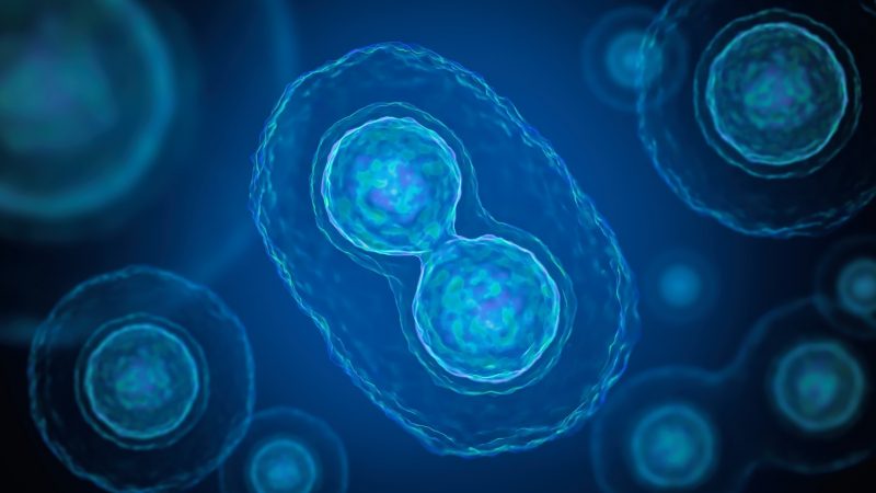
In the earliest stages of life, when an organism is still an embryo, change can happen in the blink of an eye. A single fertilised cell divides again and again as its descendants shift position, change shape, and begin taking on distinct developmental roles. Some of these transformations unfold within minutes, making the finer details extremely difficult to capture as they happen.
For scientists, the challenge is not only to see the cells clearly, but to catch them in the act. In a study published in Communications Biology, Professor Zhao Zhongying from the Department of Biology and collaborators from institutions in Hong Kong, Chinese Mainland, and the United States have developed a computational framework that brings scientists closer to doing just that.
Named EmbSAM, the framework combines a deep-learning algorithm that helps locate cell boundaries with the Segment Anything Model (SAM), a computer-vision model designed to identify and outline objects within images. Together, they allow researchers to reconstruct the three-dimensional shapes of individual cells and follow how those shapes change over time.
To put EmbSAM through its paces, the team turned to Caenorhabditis elegans, a tiny transparent roundworm that has long been a favourite of developmental biologists. Its evolution follows a remarkably consistent course and has been mapped in detail, allowing scientists to detect when individual cells divide, where they move, and what they eventually become. That makes it an ideal model for exploring how order gradually emerges from a single cell.
Watching that process, however, is easier said than done. Fluorescence microscopy allows researchers to make structures such as cell membranes glow, revealing the outlines of individual cells inside a developing embryo. But stronger laser light can bleach the fluorescent markers and damage living cells. Dial it down, and the embryo fares better, but the resulting images become noisier and the boundaries between cells harder to distinguish.
EmbSAM was designed to first enhance the weak fluorescence signal and identify where individual cells are likely to be, and then use those cues to guide SAM in tracing their boundaries across successive two-dimensional microscope images. Pieced together, these slices form complete 3D cell shapes. When tested against several existing segmentation methods, EmbSAM produced reconstructions that more closely matched manually annotated cells and remained comparatively reliable even as image noise increased.
The team then applied the framework to embryos imaged every ten seconds, bringing some of the development’s most fleeting movements into view. Cells could be seen rounding out as they prepared to divide, stretching into dumbbell-like forms and eventually separating into two daughter cells. And in the crowded confines of an embryo, no cell acts entirely alone. As dividing cells changed shape, they pushed against and deformed their neighbours, revealing the mechanical give-and-take taking place as the embryo reorganised itself.
The researchers could also follow subtle shifts in the direction of cell division as the embryo began establishing its basic body axes. Rather than jumping from one snapshot to the next, they could trace how cell shape, position and orientation changed throughout these brief yet impactful moments.
“What excites us is the possibility of moving from simply observing development to understanding the rules that shape it,” said Professor Zhao. “The more closely we can follow how cells respond to one another, the better we can begin to piece together how an embryo organises itself over time.”
The team has made the reconstructed 3D cell-shape data publicly available for further exploration, and sees potential for EmbSAM to be adapted to fluorescence imaging data from other organisms, including fruit flies, zebrafish and mice.
By bringing more of those once-missed moments into view, EmbSAM offers scientists something more valuable than a sharper image: a closer look at how form emerges from motion, and how, cell by cell, life begins to take shape.
Full article on Communications Biology: https://www.nature.com/articles/s42003-025-09220-3
Professor Zhao’s research profile: https://scholars.hkbu.edu.hk/en/persons/ZYZHAO

Professor Zhao Zhongying
Faculty of Science and Technology