
AI model learns to spot brain abnormalities without being taught what to look for
When doctors examine brain MRI scans, they look for anything unusual—whether it's a technical problem with the image itself or an unexpected sign of disease. But with the growing number of scans being performed, having an AI assistant that can automatically flag potential issues would be invaluable.
Professor GUO Xiaoqing from the Department of Computer Science, in collaboration with researchers at the University of Oxford, developed IterMask3D, an AI model that learns what a “normal” brain scan looks like by studying thousands of healthy brain images. Once trained, it can examine new scans and identify anything that deviates from normal patterns—without ever being explicitly taught what abnormalities look like.
The model works by playing a clever game of imagination. It covers different parts of the scan with digital masks and tries to “fill in” what should be underneath. If the AI cannot reconstruct certain regions because they contain patterns it has never seen during training, those areas are flagged as potentially abnormal and brought to the clinician's attention.
This approach offers several practical benefits. During the scanning process, it could detect image artifacts caused by patient movement or equipment issues (Figure 1), allowing technicians to rescan before the patient leaves. In research settings, it serves as a quality control safeguard, catching problematic images that might otherwise compromise study results (Figure 1). Perhaps most excitingly, early experiments suggest the technology may help detect brain lesions that could be overlooked during routine review (Figure 2).
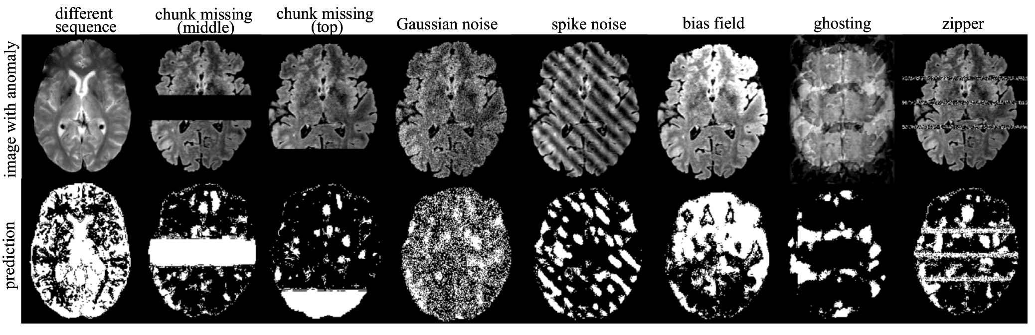
Figure 1: Visualisation of anomaly detection results on images with synthetic anomalies using our method IterMask3D. The first row shows the input images with simulated anomalies, while the second row displays the corresponding detected anomaly areas.
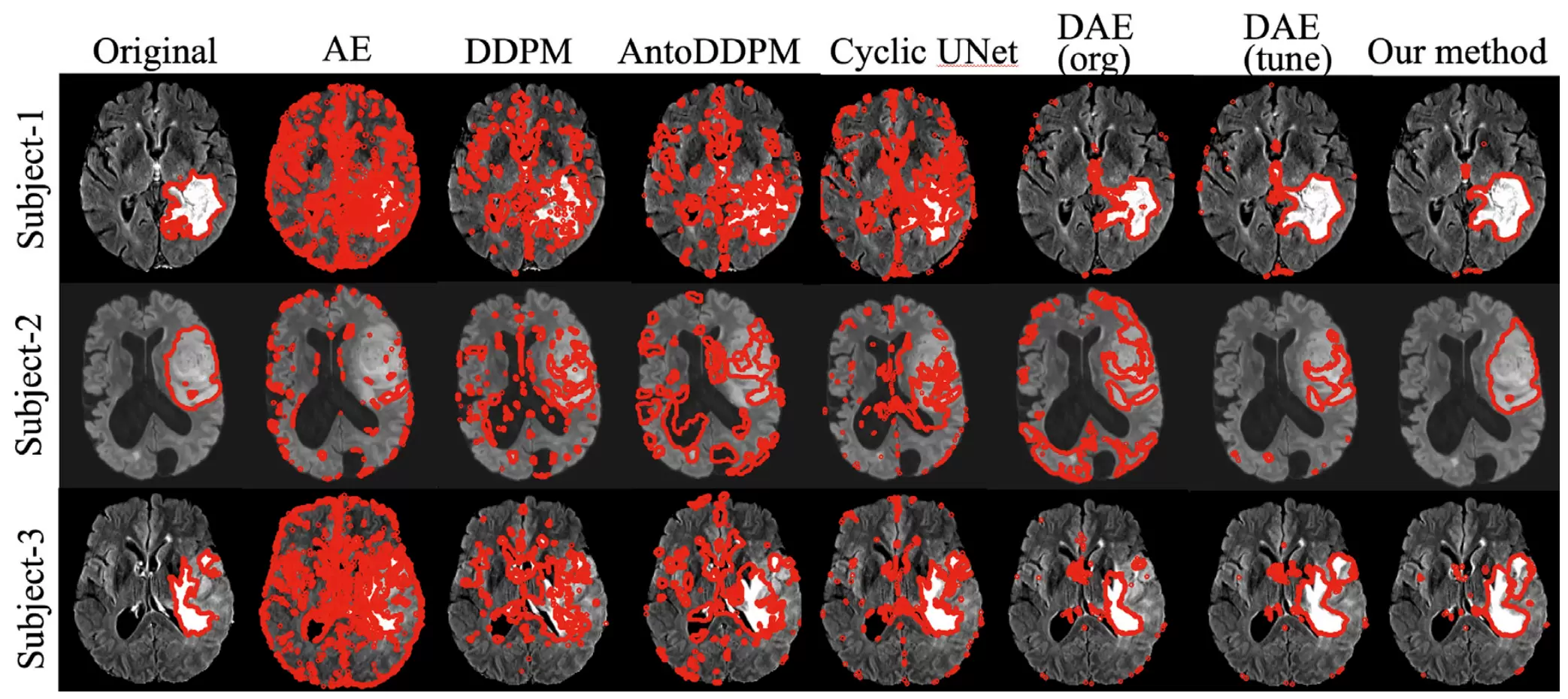
Figure 2: Anomaly segmentation results on 3D FLAIR inputs from BraTS using IterMask3D. Red contours indicate the boundaries of the predicted segmentation masks overlaid on the original images.
What makes IterMask3D particularly promising is that it does not require experts to manually label thousands of examples of different abnormalities—a process that is both time-consuming and expensive. By learning only from normal scans, this unsupervised approach can potentially detect any type of anomaly, not just those it was specifically trained to find.
The research findings have been published in Medical Image Analysis (MedIA) under the title “IterMask3D: Unsupervised anomaly detection and segmentation with test-time iterative mask refinement in 3D brain MRI”. The work was honoured with The MICCAI MedIA Best Paper Award, which recognises the highest quality paper in the Special Issue on the main MICCAI conference published in the MedIA journal.
Full research: IterMask3D: Unsupervised anomaly detection and segmentation with test-time iterative mask refinement in 3D brain MRI - ScienceDirect
More about Professor Guo's research profile: Xiaoqing GUO - Hong Kong Baptist University

Professor Guo Xiaoqing
Department of Computer Science
This article was originally published by the Faculty of Science.